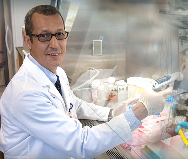
Laboratory of Azeddine Atfi, Ph.D.

Leader, Cancer Biology Program
NCI-designated Massey Cancer Center
Professor and Chair
Cellular and Molecular Pathogenesis Division
Department of Pathology
Virginia Commonwealth University
Research projects
Our research programs are dedicating to understanding the mechanisms underlying the pathogenesis and progression of human malignancies, with particular emphasis on pancreatic ductal adenocarcinoma (PDAC) and osteosarcoma.
PDAC is among the most lethal malignancies with a 5-year survival of less than 5%. Obstacles to improving patient outcomes include a lack of clear understanding of disease pathogenesis and progression, poor early detection tools, the inherently aggressive behavior of the tumor leading to early metastasis, and a lack of effective chemotherapeutics. To explore the underlying mechanisms of this deadly disease, we conducted an integrative genetic approach in which we combined the global Sleeping Beauty insertional mutagenesis system and genetic ablation of Tgif1, which encodes a general transcriptional co-repressor that we previously found to behave as an oncoprotein in breast cancer. Quite surprisingly, these genetic alterations culminated in the development of highly aggressive and metastatic PDAC tumors. Genetic profiling experiments revealed the presence of inactivating Sleeping Beauty insertions in the tumor suppressor gene Neurofibromatosis 1 (NF1), which encodes a general inhibitor of the Ras oncoproteins. From a translational perspective, we found that the NF1 gene is significantly mutated in human PDAC that do not harbor activating mutations in the proto-oncogene KRAS, which are deemed essential for PDAC initiation in 90% of PDAC patients. As such, these findings shed new insights into mechanistic paradigms of PDAC, for which no effective therapy is available.
Osteosarcoma is the most frequent primary malignancy of bone and the second leading cause of cancer-related death in adolescents. In patients without demonstrable metastasis at the time of diagnosis, surgical resection and adjuvant chemotherapy have resulted in long-term survival rates that approach 70%. However, for patients with metastatic or relapsed osteosarcoma, there is currently no reliable therapeutic option to provide long-term tumor control, emphasizing the urgent need for a better understanding of the disease. Interestingly, recent whole-genome sequencing studies revealed that the TGIF1 locus is disrupted in a significant proportion of human osteosarcoma. Comprehensive genetic experiments demonstrated that simultaneous deletion of the Tgif1 gene and activation of Sleeping Beauty mutagenesis in mesenchymal stem led to the development of highly metastatic osteosarcomas that display recurrent loss of p53 and p16Ink4a (p16), two suppressor genes frequently altered in human osteosarcoma. To further dissect the functional interplay between TGIF1 and p53/p16, we sought to explore the interaction between TGIF1 and Twist1, as Twist1 has been shown to restrict both p53 and p16 expression, and more crucially, the TWIST1 gene is frequently amplified or overexpressed in human osteosarcoma. We found that TGIF1 can associate with and repress Twist1 transcriptional activity, leading to p53 and p16 accumulation and an attendant suppression of osteosarcoma cell proliferation. In human osteosarcoma, loss of TGIF1 expression is associated with tumor aggressiveness, implicating TGIF1 as a potential prognostic marker and a possible target for attenuating the deregulated cell proliferation in this malignancy.
While studying the functional interaction between TGIF1 and Twist1 in vivo, we serendipitously found that conditional overexpression of Twist1 in mesenchymal stem cells caused severe muscle atrophy in mice with features reminiscent of cancer cachexia. Cancer cachexia is a debilitating syndrome that affects the vast majority of patients with advanced cancer and accounts for nearly 30% of cancer-related deaths. The lack of prognostic markers to identify patient susceptibility and effective treatment options for cachexia represent major gaps in cancer biology knowledge. A key feature of cancer cachexia is the progressive depletion of skeletal mass, which is mediated in part by two secreted factors, Activin and Myostatin. Recent landmark studies have shown that pharmacological inhibition of Activin/Myostatin signaling is sufficient to suppress cachexia and extend survival in several mouse models of cancer cachexia, raising the tantalizing possibility that targeting this pathway might represent a promising strategy to curb cachexia and associated morbidity and mortality in cancer patients.
Using several genetic mouse models of PDAC, we detected a massive increase in Twist1 expression in muscle undergoing cachexia. Mechanistically, tumor-derived Activin acts on muscle to induce expression of Twist1, which in turn drives expression of the muscle-specific ubiquitin ligases MuRF1 and Atrogin1, leading to muscle protein degradation. Twist1 also drives muscle Myostatin expression, reinforcing the hypothesis that Twist1 might function as a key component of the molecular networks that govern muscle cachexia. Of particular relevance, inactivation of Twist1 in mice, either genetically or pharmacologically, conferred substantial protection against muscle breakdown, which translated into meaningful survival benefits. Finally, we found that elevated levels of Twist1 in muscle were associated with severe cachexia in cancer patients, attesting to the clinical relevance of our finding. Based on these findings, we hypothesize that Twist1 might coordinate a feed-forward loop to sustain muscle wasting during cancer cachexia. Given its link to Activin/Myostatin signaling, we also hypothesize that developing combinatorial therapeutic strategies targeting both Twist1 and Activin/Myostatin signaling could mitigate potential drug toxicity by lowering the dose needed for each medicine and combat the development of resistance. We are currently undertaking a variety of experimental approaches to test these overarching hypotheses. We believe that a thorough characterization of this newly discovered cachexia network would open up new angles to the cancer cachexia field, both in terms of understanding its mechanistic paradigms and in terms of drug discovery.
Laboratory members
Allyn Bryan
Research Specialist
Creighton Friend
PhD Student
Eric Hurwitz
PhD Student
Qasim Khan
Research Specialist
Parash Parajuli
Instructor
Deanna Campbell Ward
Research Specialist
Key publications
Parajuli P, Singh P, Wang Z, Li L, Eraganreddi S, Ozkan S, Ferrigno O, Prunier C, Razzaque, M, Xu K and Atfi A. TGIF1 functions as a tumor suppressor in pancreatic ductal adenocarcinoma, EMBO Journal, 38:e101067 (2019).
Parjuli P, Kumar S, Loumaye A, Singh P, Eragmerdi S, Nguyen TL, Ozkan S, Razzaque M, Prunier C, Thissen JP and Atfi A. Twist1 activation in muscle progenitor cells during development or adulthood causes severe muscle loss reminiscent of human cancer cachexia. Developmental Cell, 45: 712-725 (2018).
Zhang MZ, Ferrigno O, Ohnishi M, Hesse E, Ferrand N, Prunier C, Razzaque M, Horne WC, Colland F, Baron R, Atfi A. TGIF Governs a feed-forward Network that empowers Wnt signaling to drive mammary tumorigenesis. Cancer Cell, 27: 547-60 (2015).
Prunier C, Zhang M-Z, Ohnishi M, Kumar S, Ferrigno O, Tzivion G, Levy L and Atfi A. PML-RAR drives acute promyelocytic leukaemia by inactivating the PHRF1 tumor suppressor. Cell Reports, 10: 883-890 (2015).
Ettahar A, Ferrigno O, Zhang MZ, Ferrand N, Prunier C, Bourgeade MF, Bieche I, Colland F and Atfi A. Identification of PHRF1 as a tumor Suppressor that promotes the TGF-β cytostatic program through release of TGIF-driven PML inactivation. Cell Reports, 4: 530-541 (2013).
Atfi A and Baron R. PTH battles TGF-b in bone. Nature Cell Biology, 12: 205-207 (2010).
Demange C, Ferrand N, Prunier C, Bourgeade MF and Atfi A. A model of Partnership Co-opted by the homeodomain protein TGIF and the ubiquitin ligase for effective execution of TNF- cytotoxicity. Molecular Cell, 36: 1073-1085 (2009).
Atfi A and Barron R. p53 brings a new twist to the Smad signaling network. Science Signaling, July 1, p33 (2008).
Faresse N, Colland F, Ferrand N, Prunier C, Bourgeade MF and Atfi A. Identification of PCTA, a TGIF antagonist that promotes PML function in TGF- signaling. EMBO Journal, 27:1804-1815 (2008).
Atfi A, Dumont E, Colland F, Bonnier D, L’Helgoualc’h A, Prunier C, Ferrand F, Clément B, Wewer U and Théret N. The disintegrin and metalloproteinase ADAM12 contributes to TGF- signaling through interaction with the Type II Receptor. Journal of Cell Biology, 178: 201-208 (2007).
Seo SR, Ferrand N, Faresse N, Prunier C, Abecassis L, Pessah M, Bourgeade MF and Atfi A. Nuclear retention of the tumor suppressor cPML by the homeodomain protein TGIF restricts TGF- signaling. Molecular Cell, 23: 547-559 (2006).
Atfi A, Abecassis L, and Bourgeade MF. Bcr-Abl activates the AKT/Fox O3 signalling pathway to restrict transforming growth factor--mediated cytostatic signals. EMBO Reports, 6: 985-991 (2005).
Seo SR, Lallemand F, Ferrand N, Pessah M, L'Hoste S, Camonis J and Atfi A. The novel E3 ubiquitin ligase Tiul1 associates with TGIF to target Smad2 for degradation. EMBO Journal, 23: 3780-3792 (2004).
Pessah M, Prunier C, Marais J, Ferrand N, Mazars A, Lallemand F, Gauthier JM and Atfi A. c-Jun interacts with the corepressor TG-interacting factor (TGIF) to suppress Smad2 transcriptional activity. Proceedings of the National Academy of Sciences USA, 98: 6198-6203 (2001).
View PubMed links:
http://www.ncbi.nlm.nih.gov/pubmed/?term=atfi+a